

Minor Immunodeficiencies and the Loss of Immune Control Over EBV, HHV-6 and HHV-7
Informational article. This material is not medical advice and does not replace consultation with a qualified physician.
Why “minor” immunodeficiencies matter in herpesvirus infections
Human herpesviruses types 4, 6 and 7 belong to a group of viruses that do not completely disappear from the body after primary infection. Instead, they enter a state of latency, meaning they remain inside cells in a “sleeping” or low-activity form.
Epstein–Barr virus, or EBV, is human herpesvirus type 4. HHV-6 is human herpesvirus type 6. HHV-7 is human herpesvirus type 7. All of them can persist in the body for years without causing obvious disease, as long as the immune system keeps them under control.
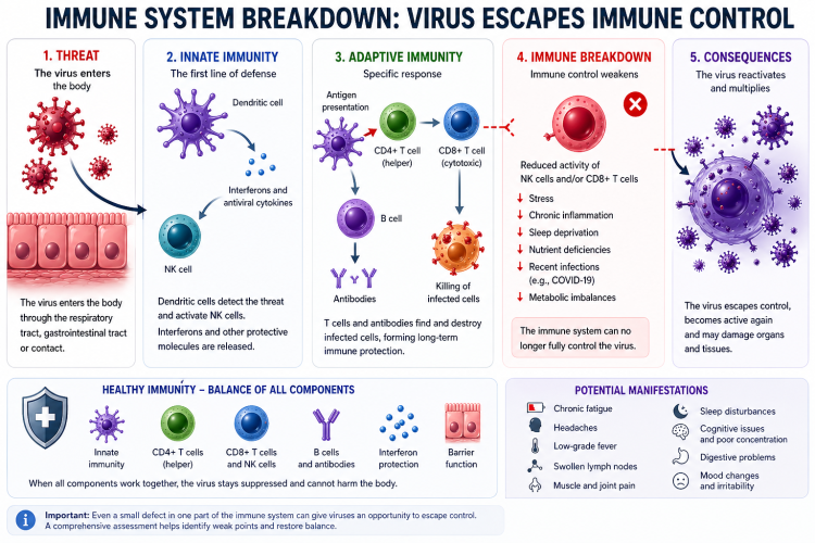
The problem begins when immune surveillance weakens. This does not always mean a severe immunodeficiency, such as HIV infection, post-transplant immunosuppression or chemotherapy. Sometimes more subtle, “minor” immune abnormalities are enough: moderate lymphopenia, reduced NK-cell activity, impaired interferon response, IgA deficiency, IgG subclass deficiency, consequences of COVID-19, severe influenza, chronic inflammation, autoimmune disease or prolonged stress.
These conditions may not look dramatic in routine blood tests, but they can reduce the body’s ability to keep herpesviruses in a latent state. As a result, the viruses may partially reactivate, sustain inflammation, worsen fatigue, low-grade fever, joint pain, lymph node enlargement, neurological symptoms and other chronic manifestations.
What is a minor immunodeficiency?
A minor immunodeficiency is not a complete “collapse” of the immune system. It is a partial, borderline or functional impairment of one or several parts of immune defense.
It may include:
- moderate reduction in lymphocytes;
- reduced CD4+ or CD8+ T cells;
- insufficient NK-cell activity;
- weak interferon response;
- IgA deficiency;
- IgG subclass deficiency;
- impaired specific antibody response;
- post-infectious immune dysregulation after COVID-19, influenza or other severe infections;
- immune dysregulation in autoimmune diseases;
- metabolic factors such as vitamin D deficiency, protein-energy malnutrition, diabetes mellitus and chronic inflammation.
Such abnormalities may be congenital, acquired or mixed. For example, a person may have a genetically weaker NK-cell response, but after COVID-19, severe stress or corticosteroid therapy this weakness may become clinically visible.
The main principle: herpesviruses are controlled by the whole immune system, not by one marker
EBV, HHV-6 and HHV-7 are not controlled by one laboratory value or one cell type. Several levels of immune defense are important:
- CD8+ T lymphocytes — cytotoxic T cells that recognize and destroy infected cells;
- CD4+ T lymphocytes — helper T cells that coordinate immune responses and support CD8+ T cells and B cells;
- NK cells — natural killer cells, cells of innate immunity that rapidly destroy virus-infected cells;
- interferons — antiviral signaling proteins, especially type I interferons and interferon-gamma;
- B cells and antibodies — important for controlling viral burden and protecting mucosal surfaces;
- mucosal immunity, especially IgA, which protects the oropharynx, respiratory tract and gut;
- complement — a system of innate immune proteins involved in recognition and elimination of infectious agents.
If one component is impaired, the body may sometimes compensate through other mechanisms. But if several moderate abnormalities are present at the same time — for example, reduced NK-cell function, IgA deficiency, persistent T-cell exhaustion after COVID-19 and chronic inflammation — herpesviruses may gain an opportunity to escape immune control.
EBV: why Epstein–Barr virus depends so much on T cells and NK cells
EBV persists mainly in B lymphocytes, especially memory B cells. Under normal conditions, the immune system continuously monitors these cells and prevents the virus from actively replicating.
The key role belongs to CD8+ T cells and NK cells. CD8+ T cells recognize viral antigens on the surface of infected cells and destroy them. NK cells act faster and less specifically: they respond to signs of cellular stress and to reduced expression of normal recognition molecules on the cell surface.
If CD8+ T cells are exhausted, if NK cells function poorly, or if the interferon response is impaired, EBV may reactivate more often. This can manifest as weakness, prolonged recovery after infections, swollen lymph nodes, tonsillitis, tonsillar plugs, low-grade fever, joint pain, cytopenias, elevated liver enzymes and, in severe cases, lymphoproliferative disorders.
HHV-6: a virus with special tropism for the immune and nervous systems
HHV-6 exists in two variants: HHV-6A and HHV-6B. HHV-6B is more commonly associated with childhood roseola, but both variants can persist in the body lifelong.
HHV-6 can infect CD4+ T cells, monocytes, macrophages, bone marrow cells and cells of the central nervous system. Therefore, its reactivation may cause not only general symptoms but also neurological manifestations: headaches, cognitive decline, severe fatigue, sleep disturbances and, in severely immunosuppressed patients, sometimes signs of encephalitis.
A special feature of HHV-6 is the possibility of chromosomal integration. This condition is called ciHHV-6 — chromosomally integrated HHV-6. In this situation, viral DNA is integrated into human chromosomes and may be detected at very high levels, without always indicating active infection. Therefore, very high and stable HHV-6 DNA levels require exclusion of ciHHV-6.
HHV-7: a less studied but potentially important virus
HHV-7, like HHV-6, belongs to the beta-herpesvirus group and has tropism for CD4+ T cells. It is widespread, is often acquired in childhood and usually remains asymptomatic.
The clinical role of HHV-7 is less well studied than that of EBV and HHV-6. Nevertheless, it has been associated with febrile illnesses, roseola-like rashes, pityriasis rosea-like skin manifestations and, in rare cases, central nervous system involvement.
One interesting mechanism of HHV-7 is its ability to evade immune recognition. One viral protein, U21, may reduce the visibility of infected cells to CD8+ T cells and NK cells. This means that even a moderate decrease in cellular immunity may create conditions for freer viral persistence.
The most important minor immunodeficiencies in EBV, HHV-6 and HHV-7 reactivation
1. Functional NK-cell insufficiency
NK cells are one of the key components of anti-herpesvirus immune control. They are especially important in the early stages of reactivation, when the virus is already awakening but the adaptive T-cell response has not yet fully activated.
The problem may involve not only the number of NK cells but also their function. Their count may be normal, while they still destroy infected cells poorly, produce too little interferon-gamma or express reduced activation receptors.
Possible markers include:
- CD16/CD56 NK cells in an immunogram;
- NK-cell cytotoxicity test;
- CD107a — a marker of NK-cell degranulation;
- perforin and granzymes;
- NKG2D, DNAM-1, NKG2A and KIR receptors — more specialized markers.
2. Moderate CD4+ lymphopenia
CD4+ T cells are coordinators of the immune response. They support CD8+ T cells, B cells and macrophages, and help form immune memory.
If CD4+ T cells are reduced or functionally exhausted, the body is less able to maintain long-term control over latent viruses. This is especially relevant for HHV-6 and HHV-7, because these viruses are closely linked to the CD4+ T-cell compartment.
Clinically, this may appear as prolonged recovery after infections, frequent viral flares, chronic fatigue, low-grade fever and unstable immunological test results.
3. Reduced CD8+ cytotoxic response
CD8+ T cells are the main cells responsible for destroying EBV-infected cells. If they are exhausted or functionally weakened, EBV gains more opportunities for reactivation.
During chronic viral stimulation, CD8+ T cells can enter a state of exhaustion. This does not mean that they disappear. They may still be present in blood, but they divide less effectively, produce fewer cytokines and destroy target cells less efficiently.
Markers of exhaustion include PD-1, TIM-3, LAG-3 and NKG2A. These are specialized parameters and are usually not included in a standard immunogram.
4. Defective interferon response
Interferons are early antiviral signaling molecules. They warn neighboring cells about a threat, slow viral replication and help properly activate NK cells and T cells.
Important components include:
- IFN-I — type I interferons, mainly IFN-α and IFN-β;
- IFN-γ — interferon-gamma, important for cellular antiviral immunity;
- IRF — interferon regulatory factors, proteins that regulate interferon responses.
After severe viral infections, including COVID-19 and influenza, the interferon system may become dysregulated: in some areas the response is insufficient, while in others it remains chronically excessive. In both situations, immune balance is disturbed.
5. Selective IgA deficiency
IgA is immunoglobulin A, the main antibody class protecting mucosal surfaces. It is important for the oropharynx, nose, airways and gut.
Selective IgA deficiency is one of the most common primary immunodeficiencies. It does not always cause severe infections, but it may increase susceptibility to chronic mucosal inflammation, recurrent respiratory infections, sinusitis, tonsillitis, gastrointestinal disturbances and autoimmune manifestations.
The connection between IgA deficiency and EBV, HHV-6 and HHV-7 reactivation is usually indirect: mucosal surfaces become inflamed more often, antigenic load is higher, infections recur more frequently and the immune system remains chronically irritated. This reduces the overall reserve of immune stability.
6. IgG subclass deficiency
IgG is the main antibody class in blood. It is divided into subclasses: IgG1, IgG2, IgG3 and IgG4. In some people, total IgG may be normal while one or more subclasses are reduced.
IgG2 is especially important for the response to bacterial polysaccharide antigens, while IgG3 often participates in antiviral responses. IgG subclass deficiency may manifest as recurrent sinusitis, bronchitis, otitis, pneumonia and prolonged recovery after infections.
In herpesvirus-related conditions, this is usually not a direct cause of reactivation, but rather a factor that maintains chronic infectious burden and exhausts the immune system.
7. Deficiency of specific antibody response
Sometimes IgG, IgA and IgM levels are normal, but the body forms poor specific antibodies after vaccination or infection. This is called impaired specific antibody response.
Such a patient may have a “normal immunogram” but still experience frequent bacterial and viral infections. Assessment may include antibody responses to vaccine antigens, such as pneumococcus, tetanus or diphtheria, depending on the clinical context.
8. Hypomorphic inborn errors of immunity
IEI means inborn errors of immunity — genetically determined immune abnormalities. They do not always manifest in childhood as severe infections. Some forms may be mild, partial and become clinically noticeable only in adulthood.
For EBV, defects affecting T-cell and NK-cell cytotoxicity are especially important. These include abnormalities in genes such as MAGT1, CD27, CD70, CTPS1, GATA2, MCM4, RASGRP1 and others. This is no longer a basic diagnostic level, but the field of clinical immunology and immunogenetics.
Such defects should be suspected in severe, unusual, familial or recurrent EBV scenarios: chronic active EBV infection, hemophagocytic syndrome, lymphoproliferation, unusual tumors, severe cytopenias or severe reactions to primary EBV infection.
Secondary immunodeficiencies after COVID-19
COVID-19 has become one of the most studied models of post-infectious immune dysregulation. In some patients after SARS-CoV-2 infection, signs of disturbed immune balance may persist: lymphopenia, T-cell exhaustion, reduced NK cells, elevated inflammatory cytokines, autoantibodies and impaired interferon response.
In Long COVID, EBV and HHV-6 reactivation are often discussed. Different studies have found increased frequency of EBV reactivation, signs of HHV-6 activity and associations with fatigue, cognitive symptoms, low-grade fever and inflammatory profiles.
It is important to understand that this does not mean EBV or HHV-6 are always the single cause of Long COVID. A more accurate interpretation is that, in some patients, post-COVID immune dysregulation creates conditions in which latent herpesviruses may reactivate and amplify symptoms.
Secondary immunodeficiencies after influenza
After influenza, a period of immune vulnerability is also possible. Post-influenza susceptibility to bacterial complications is well known: sinusitis, otitis and pneumonia. This is linked to mucosal damage and impaired function of macrophages, neutrophils, NK cells and T cells.
The connection between influenza and herpesvirus reactivation is less well studied than the connection with COVID-19. However, the mechanism is biologically plausible: severe viral infection causes systemic inflammation, temporary lymphopenia, interferon imbalance, barrier damage and exhaustion of antiviral responses. All of this may reduce control over EBV, HHV-6 and HHV-7.
Therefore, if prolonged weakness, low-grade fever, headaches, swollen lymph nodes, joint pain, tonsillitis or cognitive decline appear after severe influenza, it is reasonable to consider not only “slow recovery” but also possible post-infectious immune dysregulation with reactivation of latent viruses.
Autoimmune diseases and herpesviruses
Autoimmune diseases are themselves states of immune dysregulation. In these conditions, the immune system is not simply “weak” or “strong” — it is incorrectly tuned. Some pathways may be overactive, while others are functionally exhausted.
EBV has long been discussed in connection with systemic lupus erythematosus, multiple sclerosis, rheumatoid and other autoimmune processes. HHV-6 is also studied in the context of neuroinflammation, demyelination, chronic fatigue and post-infectious syndromes.
A vicious circle is possible: viral reactivation increases inflammation and autoimmune activity, while autoimmune inflammation and immunosuppressive therapy reduce control over viruses.
Drug-induced immunosuppression as a risk factor
Some medications may reduce immune surveillance over herpesviruses. The risk depends on dose, duration, drug combinations and the patient’s baseline condition.
Important risk factors include:
- systemic glucocorticosteroids, especially at medium and high doses;
- cytostatic drugs;
- methotrexate in susceptible patients;
- anti-CD20 therapy, such as rituximab;
- JAK inhibitors;
- some biologic drugs used in autoimmune diseases;
- post-transplant immunosuppressive therapy.
This does not mean these medications cannot be used. But in a patient with a history of EBV, HHV-6 or HHV-7 reactivation, it is preferable to understand the viral and immune background in advance.
Why routine tests often fail to show the problem
A routine complete blood count may be almost normal. Immunoglobulins may remain within reference ranges. IgG antibodies to EBV, HHV-6 or HHV-7 are positive in most adults and do not, by themselves, prove active infection.
The problem is that a minor immunodeficiency is often functional. It may appear not as a sharp drop in one marker, but as a weak response under immune stress.
For example:
- lymphocytes may be at the lower end of normal;
- CD8+ T cells may be present in sufficient numbers but functionally exhausted;
- NK cells may be sufficient in number but have reduced cytotoxicity;
- total IgG may be normal, while IgG3 is reduced or the specific antibody response is impaired;
- viral PCR may be weakly positive while symptoms are significant;
- serology may be ambiguous.
Therefore, when such a problem is suspected, one “magic” test is not enough. Clinical features, immunogram, virological testing and symptom dynamics must be interpreted together.
How to interpret positive herpesvirus tests correctly
Positive IgG antibodies to EBV, HHV-6 or HHV-7 usually mean past exposure, not active disease. In most adults, these antibodies are positive.
For EBV, the pattern is more important: VCA IgM, VCA IgG, EBNA IgG, EA IgG and, when an active process is suspected, quantitative EBV DNA PCR.
For HHV-6 and HHV-7, serology is less informative. Quantitative PCR is more important, but it must be interpreted carefully: the sample type, viral load, repeatability of the result, clinical picture and immune background all matter.
Most importantly, the presence of viral DNA without symptoms and without immunological abnormalities does not always require treatment. But viral DNA plus a compatible clinical syndrome plus an immune defect is a much more significant combination.
Main clinical conclusion
Reactivation of EBV, HHV-6 and HHV-7 is rarely explained by one single cause. More often, it is the result of several overlapping factors: congenital predisposition, previous COVID-19 or influenza, chronic inflammation, mucosal immune deficiency, functional NK-cell weakness, T-cell exhaustion, autoimmune activity, stress, sleep deprivation and metabolic abnormalities.
This is why a patient may remain in a “gray zone” for years: formally, there is no severe immunodeficiency, but the immune system no longer holds latent viruses under perfect control. This is the area of minor immunodeficiencies — subtle, partial but clinically meaningful disturbances of immune balance.
What should not be missed
If, after COVID-19, influenza, severe stress or an autoimmune flare, a person develops prolonged fatigue, low-grade fever, lymph node inflammation, headaches, joint pain, frequent tonsillitis, tonsillar plugs, persistent sinusitis, skin manifestations, impaired concentration and unclear systemic symptoms, it is worth considering not only a single infection but the state of immune control as a whole.
In such a situation, the most useful approach is not to search for one virus as the only cause, but to assess the whole combination: viral load, clinical syndrome, T cells, NK cells, antibodies, mucosal immunity, inflammation, autoimmune markers and metabolic background.
This comprehensive approach helps distinguish an incidental laboratory finding from clinically significant reactivation and identify the main point of intervention: antiviral therapy, restoration of immune balance, correction of deficiencies, treatment of autoimmune disease or reduction of chronic inflammatory burden.
Informational article. This material is not medical advice and does not replace consultation with a qualified physician.
 АС
АС